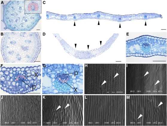
Figure 2.
Comparison of Internal Structures and Epidermal Cell Shapes of the Stem, Axillary Shoot, Cladode, and Scale Leaf in A. asparagoides.
(A) Mature stem. Inset shows close-up view of the vascular bundle; top of the image is the side facing the stem center.
(B) Axillary stem.
(C) and (D) A part of a cladode (C) and a scale leaf (D).
(E) Close-up view of the inner structure of a cladode.
(F) and (G) Close-up views of the vascular bundle of a scale leaf (F) and of a cladode (G).
(H) to (M) Scanning electron micrographs of a stem epidermal cell (H), axillary shoot (I), adaxial side of a scale leaf (J), abaxial side of a scale leaf (K), adaxial side of a cladode (L), and abaxial side of a cladode (M).
(A) to (G) are transverse sections. In (C) to (G), top of the image is the adaxial side of each organ. In (A), (F), and (G), the xylem is colored in pink. Black and white arrowheads indicate vascular bundle and stoma, respectively. p, phloem; x, xylem. Bars = 1 mm in (C) and (D) and 100 μm in (A), (B), and (E) to (G).

