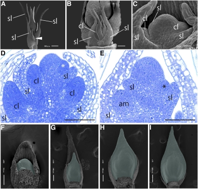
Figure 3.
Development of Cladodes in A. asparagoides.
(A) to (C) Scanning electron micrographs of the shoot apex.
(A) Axillary shoots arising on the axils of primary shoot scale leaves. White arrowhead indicates the axillary shoot.
(B) Close-up view of an axillary shoot meristem composed of scale leaves and cladode primordia.
(C) Close-up view of cladodes arising at the axil of scale leaves on the axillary shoot meristem.
(D) Longitudinal section of the axillary shoot meristem with emerging cladodes. White asterisk indicates the emerging cladode.
(E) Longitudinal section of the primary shoot with emerging axillary shoot meristem. Black asterisk indicates the emerging axillary shoot meristem.
(F) to (I) Scanning electron micrographs of cladode development in series. Cladodes are colored green.
Unnecessary organs were removed in (B), (C), and (F) to (I). am, axially shoot meristem; cl, cladode; sl, scale leaf. Bars = 100 μm in (D) and (E).

