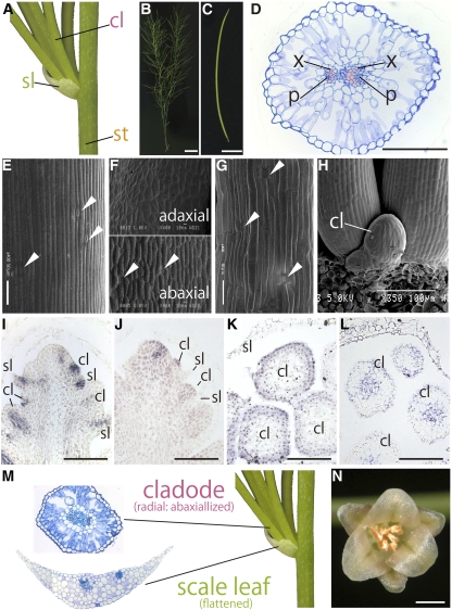
Figure 7.
Morphology, Anatomy, and Ortholog Expression Patterns Associated with Shoot and/or Leaf Morphogenesis Genes in A. officinalis.
Generating position of cladodes (A), gross morphology of a shoot (B), and cylindrical cladode of A. officinalis (C).
(D) Internal structure of the cylindrical cladode. Top of the image is the adaxial side of the cladode.
(E) to (G) Scanning electron micrographs of epidermal cells from the stem (E), scale leaf (F), and cladode (G).
(H) Close-up view of arising cladodes.
(I) to (L) In situ localization of Aof AS1 and Aof KNAT1 transcripts in an axillary shoot bearing leaf and cladode primordia and of miR166 and Aof PHB transcripts in cladode primordia. Aof AS1 transcripts (I) and Aof KNAT1 transcripts (J) in a longitudinal section of an axillary stem subtending leaf and cladode primordia. In situ localization of miR166 (K) and Aof PHB transcripts (L) in a transverse section of cladode primordia.
(M) Comparison of the internal structure of an A. officinalis cladode and scale leaf.
(N) Gross morphology of an A. officinalis male flower.
In (D), the xylem is colored pink. cl, cladode; p, phloem; sl, scale leaf; st, stem; x, xylem. White arrowhead indicates stoma. Bars = 1 cm in (B), 0.5 cm in (C), 100 μm in (D) and (I) to (L), and 1 mm in (N).

