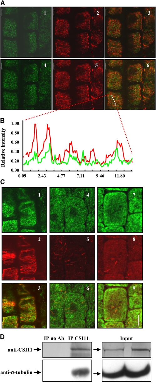
Figure 6.
CSI1 Binds to Microtubules in Vivo.
(A) Colocalization of CSI1 and cortical microtubules in Arabidopsis root cells. Immunolocalization of CSI1 using anti-CSI1 antibody (in green, [1] and [4]) and anti-α-tubulin–labeled microtubules (in red, [2] and [5]) are merged (3) and (6). Single image shot (1) to (3) and projection of 10 images at an interval of 0.2 μm (4) to (6) are shown.
(B) Intensity of a plotted line by scanning through a merged region of CSI1 using anti-CSI1 antibody and anti-α-tubulin–labeled microtubules (image 6 of [A], the scanned region is highlighted by a dashed line) confirmed the correlation between CSI1 and microtubules.
(C) Localization of CSI1 after oryzalin-induced microtubule depolymerization. Immunolocalization of CSI1 using anti-CSI1 antibody (in green, [1], [4], and [7]) and anti-α-tubulin–labeled microtubules (in red, [2], [5], and [8]) are merged (3), (6), and (9). Oryzalin treatment (10 μM) for 0 h (1) to (3), 0.5 h (4) to (6), and 4 h (7) to (9) are shown.
(D) Coimmunoprecipitation of CSI1 and α-tubulin. Four-week-old Arabidopsis plant extracts were immunoprecipitated with protein G conjugated beads preincubated with or without rabbit anti-CSI1 antibody, followed by SDS-PAGE and immunoblot using anti-CSI1 or goat anti-α-tubulin. Immunoprecipitation of CSI1 and coimmunoprecipitation of α-tubulin are shown. No cross-reactivity to eluted antibody at 25 kD or 50 kD was detected. Empty beads without antibodies were used as a negative control, and expression of CSI1 and α tubulin in both input extracts was examined (right panel). Ab, antibody; IP, immunoprecipitation.
Bars in (A) and (B) = 5 μm.

