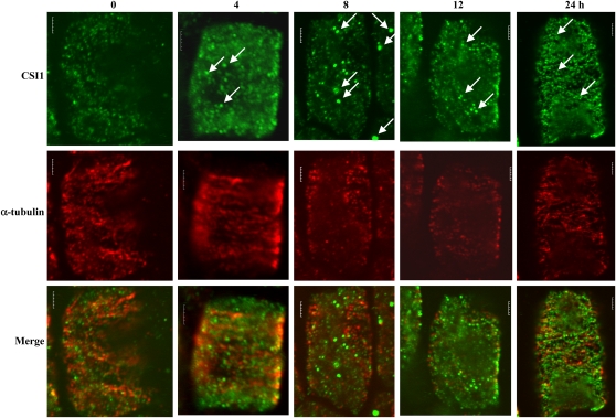
Figure 9.
Dynamic Change of CSI1 and Microtubules under Dehydration Treatment.
Immunolocalization of CSI1 using anti-CSI1 antibody (green) and anti-α-tubulin–labeled microtubules (red) of wild-type root cells exposed to treatment with PEG6000 (20%, w/v, in liquid MS media) for 0, 4, 8, 12, and 24 h, respectively. Arrows indicate the induced aggregation of CSI1 after dehydration treatment.
Bars = 5 μm.

