
Figure 1.
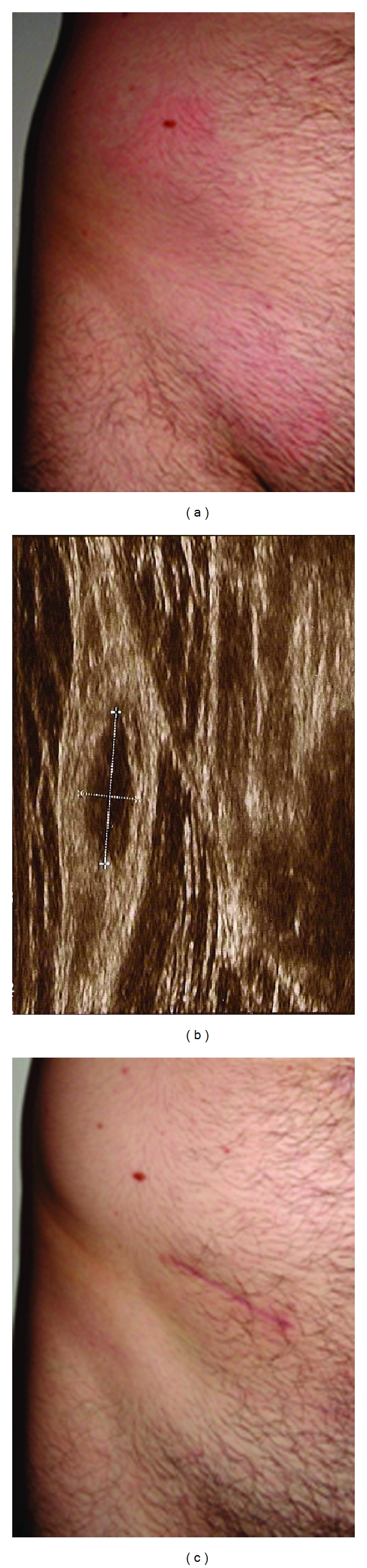
Clinical picture before and after surgery. Before the removal of the parasite, the patient showed erythematous plaques with mild edema in the lower right abdominal region (a). The ultrasound revealed a well-defined cystic lesion of 11.9 mm × 4.8 mm above muscular layer, which is seen on the right (b). After excision of the nodule the wound healed without complications and all symptoms resolved. The scar reveals the final location of the parasite (c).