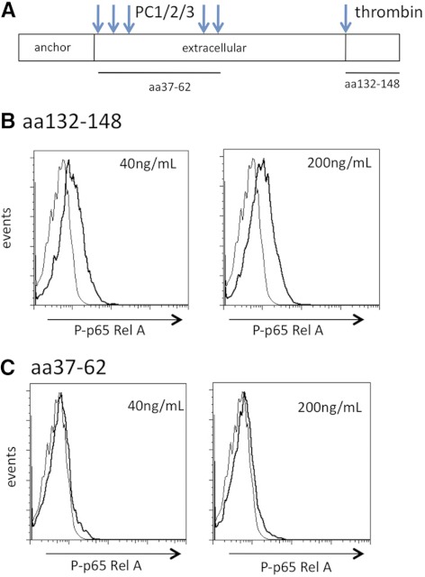
Figure 7. Ecrg4 CΔ16 peptide induced the P-p65.
(A) Diagram of Ecrg4 peptides tested for macrophage activation. (B) Effect of the Ecrg4 CΔ16 peptide (SPYGFRHGASVNYDDY) on the P-p65 in primary peritoneal mouse macrophages incubated at 40 ng/mL and 200 ng/mL (20 nM and 100 nM, respectively) for 15 min at 37°C. Representative plots are shown (n=3). (C) Effect of Ecrg4 37–62 (MLQKREAPVPTKTKVAVDENKAKEFL) on P-p65 in macrophages incubated with 40 ng/mL and 200 ng/mL (15 nM and 70 nM, respectively; n=3).

