Figure 2.
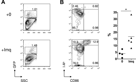
Effect of topical imiquimod on LC frequency and phenotype in the skin epithelium. (A) Contour plots showing GFP reporter expression (specific for host LCs) in skin epithelia of Langerin.DTR/GFP mice treated 24 hours previously with topical imiquimod (Imq) or control. (B) Left: Contour plots showing dual staining for CD86 and MHC class II in gated LCs from mice treated 24 hours previously with topical imiquimod or control. Right: Summary data showing the percentage of CD11b+ LCs that were CD86high MHCIIhigh, in control or imiquimod-treated mice (n = 4 or 5 mice per group); data pooled from 3 independent experiments. *P < .05. The mean is indicated by the horizontal bar.

