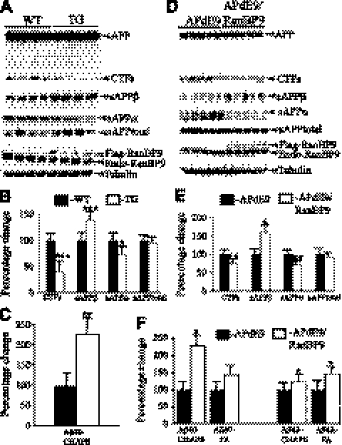
Figure 2.
RanBP9 overexpression increases amyloidogenic processing of APP in vivo. A–C) APP metabolites measured in 4-mo-old WT and RanBP9-transgenic (TG) mice. A) Lysates prepared from cortical tissue using Nonidet P-40 buffer were subjected to immunoblotting and probed with specific antibodies. B) RanBP9 overexpression decreased levels of CTF by 60%, sAPPα by 28%, but sAPPβ levels were increased by 40%, while sAPPtotal levels were not altered (n=4/group). C) CHAPSO-soluble Aβ40 was extracted from hemibrains and quantified by ELISA. RanBP9 overexpression in APdE9/RanBP9 triple-transgenic mice (n=4, age 4 mo) increased Aβ40 levels by 122% compared to APdE9 double-transgenic mice (n=4, age 4 mo). D–F) APP metabolites in 12-mo-old APdE9 and APdE9/RanBP9 mice. D) Cortical lysates were subjected to immunoblotting for quantification of APP metabolites. E) RanBP9 overexpression in APdE9 mice decreased levels of CTF by 24%, sAPPα by 29%, but sAPPβ levels were increased by 59%, while sAPPtotal levels were not altered (n=5/group, age 12 mo). F) CHAPSO-soluble and FA-soluble Aβ was extracted from hemibrains and quantified by ELISA (n=6/group, age 12 mo). Aβ40 was increased in both CHAPSO-soluble and FA-soluble fractions, while Aβ42 was increased only in FA-soluble fractions. Aβ was quantified per milligram protein. Values are expressed as percentage change from APdE9 control. Data are means ± se. *P < 0.05, **P < 0.01, ***P < 0.001 vs. control; Student's t test.

