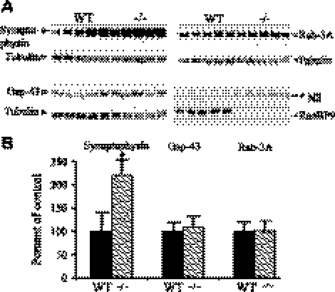
Figure 9.
Neonatal RanBP9-null mice showed increased presynaptic proteins. A) Brain homogenates from RanBP9-null mice (−/−) and age-matched WT controls prepared using Nonidet P-40 lysis buffer were subjected to SDS-PAGE electrophoresis and probed with antibodies against presynaptic proteins, synaptophysin, gap-43, and Rab-3A. Monoclonal RanBP9 antibody detected endogenous RanBP9 protein only in WT mice, while RanBP9-null mice showed complete absence of RanBP9 protein. *NS, nonspecific band. B) ImageJ quantification showed that synaptophysin was increased by 122%, but gap-43 and Rab-3A levels were not altered. Data are presented as means ± se (n=5/group). *P < 0.05 vs. WT; Student's t test.

