Figure 2.
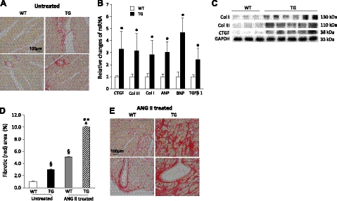
ROCKΔ1 transgenic mice developed interstitial and perivascular fibrotic cardiomyopathy. A) Heart sections were stained with Sirius red. Transgenic (TG) heart showed noticeable collagen deposition (red) in interstitial and perivascular regions compared with the wild-type (WT) heart under physiological (untreated) conditions. B, C) Significant increases in fibrotic and stress factors were detected in the transgenic hearts by qPCR (B) and immunoblot analyses (C). qPCR data were pooled from 10 mice/group with analyses for each mouse in triplicate. D) Quantification of fibrotic areas. E) Even more severe fibrotic ardiomyopathy was observed with Sirius red staining in the mutant heart after ANG II treatment compared with the WT heart. col, collagen. *P < 0.01 vs. WT; **P < 0.01 vs. ANG II-treated WT; §P < 0.01 vs. untreated WT; ΔP < 0.01 vs. untreated TG.

