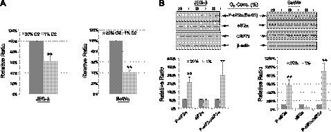
Figure 4.
Hypoxia reduces trophoblast cell proliferation and induces eIF2α phosphorylation. A) Both JEG-3 and BeWo cells were cultured at either 20 or 1% O2 in normal culture medium for 3 d. Number of cells was counted using a hemocytometer. Experiments were done in duplicate and repeated 3 times. Data presented are means ± sd; the value of 20% O2 is used as a control and expressed as 100%. B) Top panel: proteins were isolated from both JEG-3 cells and BeWo cells after 3 d incubation and subjected to Western blot analysis. Anti-P-eIF2α, anti-eIF2α, and anti-GRP78 antibodies were used. β-Actin was used as a loading control. Bottom panel: densitometric quantification of band intensity. Levels of phosphorylated and total protein are presented as a ratio to the untreated control (100%). Data are means ± sd from 3 independent experiments. **P < 0.01.

