Figure 4.
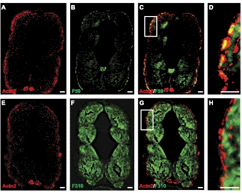
α-Actinin-2 is expressed in slow fibers in zebrafish skeletal muscle. A, B) Immunofluorescence was performed on the transverse sections of wild-type zebrafish (5 dpf) with α-actinin-2 antibody (F6523) against zebrafish protein (A) or F59 antibody that strongly labels superficial layer of slow muscle cells and weakly labels deep fast muscle cells (B). C) Immunolabeling with both F6523 and F59 antibody showed colocalization of 2 proteins (yellow signal). D) High-magnification view of boxed area in C. E–H) In contrast, immunolabeling with fast muscle marker F310 did not show any colocalization with F6523. E) F6523 antibody. F) F310 antibody. G) F6523 and F310 antibody. H) High-magnification view of boxed area in G. Scale bars = 30 μm (A–C, E–G); 10 μm (D, H).

