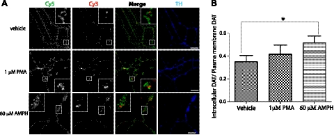
Figure 6.
Quantitative analysis of HA-DAT endocytosis in axons. A) HA11 antibody-feeding endocytosis assays were carried out as described in Materials and Methods. Postnatal cultures were incubated with HA11 antibody at 37°C for 4 h, washed, and then incubated with vehicle (DMSO), 1 μM PMA, or 60 μM AMPH at 37°C for 1 h. Cells were then fixed and stained with Cy5-conjugated secondary antibody to label the plasma membrane HA-DAT (green), followed by permeabilization and staining with Cy3-conjugated secondary to label internalized HA-DAT (red). Neurons were also stained with the TH antibody (blue). Scale bars = 10 μm. B) Quantification of the Cy3/Cy5 ratio from experiments exemplified in A was performed as described in Materials and Methods. Bars represent means ± se (n=14). Similar results were obtained in ≥4 independent experiments. *P < 0.05.

