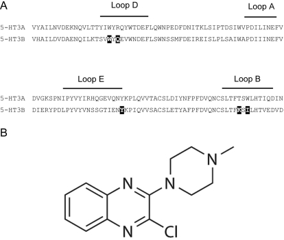
Fig. 1.
A, an alignment of 5-HT3A and 5-HT3B subunit residues. B subunit residues that were Cys-substituted in this study are shown as white text on a black background. The effects of these residues on 5-HT activation and [3H]granisetron binding have already been studied elsewhere (Thompson et al., 2011b), and each aligns with an A subunit residue that has been shown to be an important binding site residue (for review, see Thompson et al., 2010a). B, structure of VUF10166.

