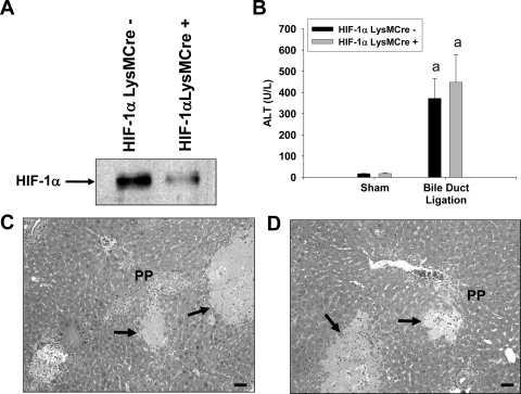
Fig. 7.
Liver injury in HIF-1αLysMCre− and HIF-1αLysMCre+ mice after BDL. A, Kupffer cells were isolated from HIF-1αLysMCre− and HIF-1αLysMCre+ mice and exposed to 1% oxygen for 1 h. HIF-1α was detected in nuclear extracts by Western blot analysis. HIF-1αLysMCre− and HIF-1αLysMCre+ mice were subjected to sham operation or BDL. B, 10 days after surgery, ALT activity was measured in serum. Data are expressed as mean ± S.E.M.; n = 8. a, significantly different (p < 0.05) from sham-operated mice. C and D, representative photomicrographs from HIF-1αLysMCre− (C) and HIF-1αLysMCre+ (D) mice subjected to BDL. Arrows indicates area of necrosis. Scale bar, 50 μm.

