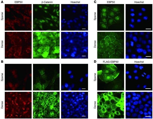
Figure 1. EBP50 displays nuclear translocation under low-density culture.
SW480 (A) and HT29 (B) colon cancer and MDCK (C) cell lines were plated in either sparse or dense conditions for 2 days and processed for EBP50 and β-catenin immunofluorescence study as well as Hoechst 33342 nuclear staining. (D) MDCK cells stably expressing FLAG-tagged EBP50 were processed for FLAG and Hoechst 33342 nuclear staining. Scale bars: 10 μm.

