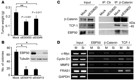
Figure 7. Tumorigenesis of the colon cancer cells is reduced by EBP50 knockdown, which disrupted β-catenin and TCF-1 interaction.
(A) Mock and EBP50 siRNA stably transfected (siE50) SW480 clones were injected beneath the dorsal skin of 6-week-old NOD-SCID mice. After 3 weeks, the mice were sacrificed. The injected tumors (n = 5 for each group) were dissected from the mice and weighed, and the results are shown (mean ± SEM). (B) Mock and EBP50 siRNA stably transfected HT29 clones were processed for soft agar assay to assess their anchorage-independent growth ability. The histogram shows results (mean ± SEM) from 3 different fields for each clone. The inset shows the result from Western blot analysis of EBP50 in mock and siRNA-expressing clones. *P < 0.05, **P < 0.001. (C) Cell lysates from mock and siE50 SW480 clone were immunoprecipitated with equal amounts of mouse normal serum (Ctr) and anti–β-catenin monoclonal antibody and processed for Western blot analysis using the indicated antibodies. (D) Mock (M) or siEBP50 SW480 (Si) cells were processed for ChIP assay using rabbit anti-EBP50, anti–β-catenin, or anti–TCF-1 antibody. Five percent of the immunoprecipitated DNA served as the templates for amplifying all the target sequences by PCRs, except those for MMP2, which used 0.5% of the immunoprecipitated materials as the templates for PCRs.

