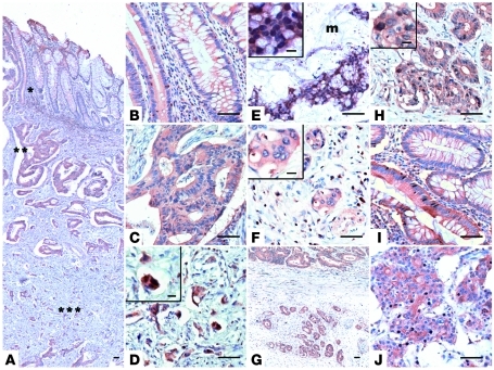
Figure 8. EBP50 expression in colorectal cancer specimens.
A representative CRC sample showed global EBP50 staining (A) in the mucosa (*), submucosa (**), and deeper invasive portions of the tumor (***); these regions are presented as magnified images in B–D, respectively. (E) EBP50 expression in CRC specimen with extracellular mucin pooling (m). (F) A representative sample showing negative nuclear EBP50 staining in the invasive front. Low- (G) and high-magnification (H) images of another CRC sample, which demonstrates aberrant nuclear EBP50 expression in the peripheral tumor nests (lower portion in G) outside the main tumor mass (upper portion in G). EBP50 expression was infrequently found concomitantly in the nuclei of the well-differentiated epithelial portion (I) and the poorly differentiated cancer part (J) in only a single case among our CRC patients. Scale bars: 50 μm; 10 μm, insets in D–F and H.

