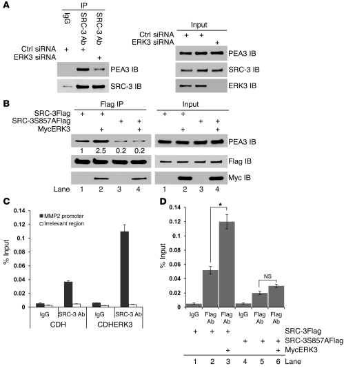
Figure 6. ERK3 promotes the interaction of SRC-3 with PEA3 and occupancy of SRC-3 on the PEA3 binding site of MMP2 gene promoter, which is mediated by the phosphorylation site S857.
(A) Knockdown of ERK3 decreases the interaction of SRC-3 with PEA3. H1299 cells were transfected with ERK3 siRNA or nontargeting control siRNA. Coimmunoprecipitation was performed using a SRC-3 Ab or a mouse IgG, followed by Western blotting. (B) ERK3 promotes the interaction of SRC-3 with PEA3, which is mediated by the phosphorylation site S857. H1299 cells were transfected with SRC-3Flag or SRC-3S857AFlag or together with Myc-tagged ERK3 (MycERK3). Coimmunoprecipitation was performed using a Flag Ab. Numbers below the PEA3 immunoblots in Flag immunoprecipitation represent the relative intensity of the protein bands. The band intensity in lane 1 is set as “1.0.” (C) Overexpression of ERK3 enhanced occupancy of SRC-3 on the MMP2 promoter. H1299 cells were stably transduced with either lentiviral vector CDH or lentiviral ERK3 (CDHERK3). ChIP assays were performed using either a SRC-3 Ab or goat IgG. SRC-3 protein occupancy on the PEA3 binding site of MMP2 gene promoter (700-bp upstream of transcription start site; ref. 19) was analyzed by quantitative real-time PCR and presented as the percentage of sheared chromatin input. (D) ERK3 promotes the occupancy of SRC-3, but not the SRC-3S857A mutant, on the MMP2 promoter. A549 cells were singly transduced or cotransduced stably with the lentiviral constructs as indicated. ChIP assays were performed using either a Flag Ab or mouse IgG. *P < 0.01.

