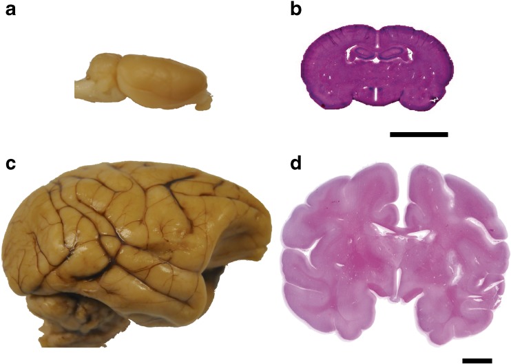
Fig. 1.
Comparison of lissencephalic and gyrencephalic brain structure. (a) The rat brain is lissencephalic; gross structure demonstrates smooth cortex with absence of sulci. (b) Histological rat brain section in coronal plane stained with hematoxylin & eosin demonstrates smooth cortical anatomy and high ratio of gray:white matter. (c) Gyrencephalic cynomolgus macaque brain; gross structure demonstrates multiple sulci and gyri. (d) Histological cynomolgus macaque brain section in coronal plane stained with hematoxylin & eosin demonstrates gyri and sulci, organized white matter tracts, subcortical nuclei, and a lower ratio of gray:white matter than rats. Histology scale bars = 10 mm

