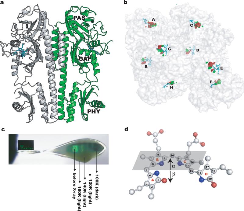
Fig. 1.
Trap-pump-trap-probe experiment. (a) Ribbon diagram of the PaBphP-PCM dimer. The BV chromophore is colored in cyan. (b) Experimental difference (Flight-Fdark) map at 130K (contoured at +/-5σ, where σ is the standard deviation of difference densities across the entire map). Strong positive (green) and negative (red) densities with peak signal greater than +/-12σ are clustered near the chromophores of the eight monomers (A-H) in the asymmetric unit. (c) Dark stripes of a mounted crystal correspond to segments from which X-ray datasets were collected. (d) A ball-and-stick representation of the chromophore in the Pfr state (PDB 3NHQ). The α-face of a pyrrole ring is defined when atom numbering follows a clockwise direction, with β defined as the opposite face. The α-face of an entire bilin chromophore is defined according to Rockwell et al.30.

