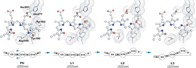
Fig. 4.
Light-induced molecular events in PaBphP. (Upper panel) The chromophores are in ball-and-stick representation, with their surrounding residues shown as van der Waals spheres. Green dotted lines indicate potential interactions with each cryo-trapped structure. (Lower panel) Schematic representation of changes in relative disposition of the four pyrrole rings of the BV chromophore, in which pyrrole rings A, B, C and D (boxes) are linearly connected by methine bridges (circles). The α- and β-faces of the chromophore are denoted by arrows.

