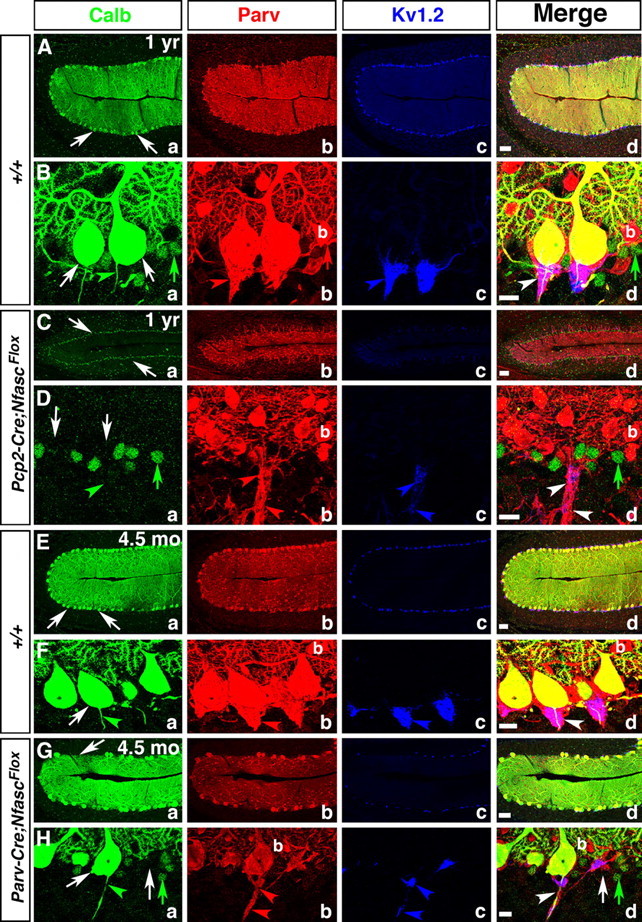
Figure 8.

Ablation of Nfasc in Purkinje neurons leads to progressive Purkinje neuron degeneration. One-year old wild-type (A, B), one-year-old Pcp2-Cre;NfascFlox (C, D), 4.5-month-old wild-type (E, F), and 4.5-month-old Parv-Cre;NfascFlox (G, H) cerebellar sections immunostained against Calb (a, green), Parv (b, red), KV1.2 (c, blue), and merged (d). One-year and 4.5 month wild-type cerebella maintain a full complement of healthy Purkinje neuron layer (Aa, Ba, Ea, Fa, arrows), while one-year-old Pcp2-Cre;NfascFlox cerebella show decreased molecular layer size with loss of Purkinje neurons (Ca, Da, arrows). The pinceau is stably maintained in one-year and 4.5-month-old wild-type cerebella (Bb–d, Fb–d, arrows). In Pcp2-Cre;NfascFlox cerebella, the basket axons appear clumped together (Db, d, arrowheads), but do not form a pinceau structure as all Purkinje neurons have died (Da, arrows). In 4.5 month Parv-Cre;NfascFlox cerebella, some Purkinje cells have begun to degenerate (Ga, d; Ha, d, arrows) and the pinceau structure remains disrupted with ectopic clusters of potassium channels (Hb–d, arrowheads). Scale bars: (in d) A, C, E, G, 50 μm; B, D, F, H, 10 μm.
