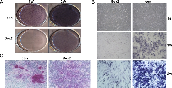
Fig. 3.
Comparison of ALP staining and mineralized nodules between Sox2 expressing cells and control cells. a ALP staining of Sox2-expressing cells and control cells. b The details of ALP staining and cell morphology change after being induced with osteogenic differentiation medium. c Alizarin red staining of Sox2-expressing cells and control cells

