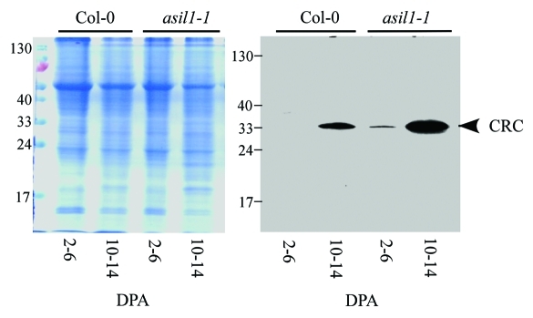
Figure 2.
Accumulation of the 12S globulin cruciferin in developing seeds of asil1 mutant. Total protein extracts were prepared from siliques of wild type (Col-0) and asil1–1 mutant plants corresponding to stages 2–6 DPA and 10–14 DPA. An equal amount of protein (10 µg) was loaded in each lane and subjected to SDS-PAGE followed by either Coomassie brilliant blue staining (left) or immunoblot analysis with monoclonal anti-12S cruciferin (CRC) antibody (right) as described previously.1

