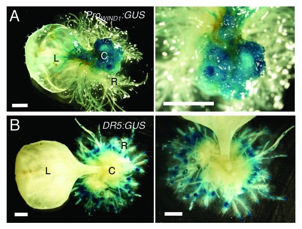
Figure 3.
WIND1 expression persists in dedifferentiated callus cells. (Left panels) Rosette leaves of 14-d-old plants carrying the ProWIND1:GUS (A) and DR5:GUS (B) constructs were cultured on 1 µM NAA containing MS medium for 30 d. (Right panels) Magnified view of the NAA-induced callus and roots. The ProWIND1:GUS signal is strong in callus cells while the DR5:GUS signal is not found in callus and detectable remarkably in roots in contact with the medium. L: leaf; C: callus; R: regenerated roots from callus. Scale bars, 1 mm (A, B)

