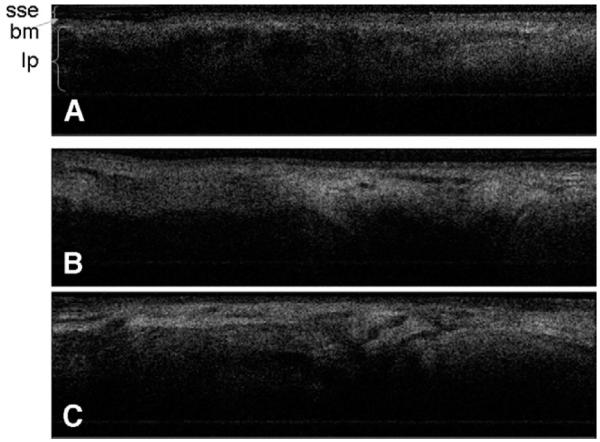
Figure 4.

OCT images demonstrate representative laryngeal subsites. (A) Left true vocal cord; (B) right true vocal cord; (C) interarytenoid mucosa, note the increased glandular structures seen in this location. (sse, stratified squamous epithelium; bm, basement membrane; lp, lamina propria.)
