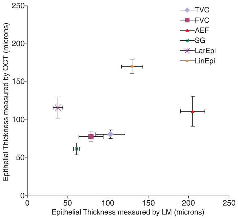
Fig. 3.
Mean epithelial thickness at each laryngeal subsite measured by light microscopy (LM) and optical coherence tomography (OCT), with standard errors. TVC, true vocal cords; FVC, false vocal cords; AEF, aryepiglottic folds; SG, sublgottis; LarEpi, laryngeal epiglottis; LinEpi, lingual epiglottis.

