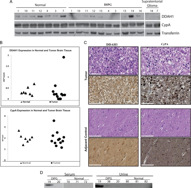
Fig. 3.
CypA and DDAH1 expression analysis using frozen brain tissue, serum, and urine specimens. (A) Western blot analysis showed equal expression levels of both CypA and DDAH1 in frozen diffuse intrinsic pontine glioma (DIPG) tumors (n = 12) and normal brain tissue (n = 7), indicating no alteration in cytosolic levels of these proteins. (B) Graphical representation of Western blot analysis of DIPG tumor tissue and control brain tissue based on densitometry of Western blots shown in panel A normalized to GAPDH expression levels. (C) Immunohistochemical staining of DIPG tumor tissue demonstrates cytosolic expression of CypA and DDAH1 in high-grade regions, as indicated by H&E staining. Adjacent normal regions as identified by a pathologist were used as controls. (Scale bar = 10 μm). (D) Western blot analysis of serum and urine specimens collected from patients with DIPG demonstrate expression of CypA in serum from patient 19 and urine of patients 19 and 38. Low expression level of CypA was detected in serum of one control patient (number 72) lacking intracranial pathology. DDAH1 was not detected in serum and urine of these patients (data not shown).

