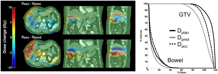
Fig 3.
Deviations from the accumulated dose (Dacc) are shown. PTV coverage was compromised inferiorly on the static exhale CT plan (Dplan) to spare the large bowel. 4D CT (Dpred) predicted less dose as these tissues moved inferior away from the high-dose region. Geometric errors seen on 4D CBCT moved these tissues back towards the high-dose region.

