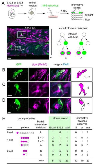
Fig. 8.
Retrovirally marked clones exhibit symmetric and asymmetric patterns of Math5 expression. (A) E12.5 or E13.5 retinas were explanted from Math5 lacZ/+ embryos, flattened on polycarbonate membranes, infected at low density with a retroviral stock to mark clonal lineages (green), and cultured for 3 days in vitro (DIV). The micrograph shows a cross-section from a representative explant (bracket) co-stained for cytoplasmic βgal (magenta) and GFP (green). The diagram shows hypothetical 2-cell clone with βgal+ cells. Each clone reflects one informative terminal division: a symmetric [S] division which gave rise to two Math5+ daughters (left); or an asymmetric [A] division, which gave rise to one Math5+ and one Math5- daughter (right). (B-D) Confocal Z-stack projections and drawings showing representative clones that are symmetric (B, C) or asymmetric (D) with respect to Math5 expression. (E) Summary of observed clones containing at least one Math5+ cell. Informative divisions have a unique interpretation, and give rise to one [A] or two [S] Math5+ daughters. Both types of divisions were identified. MIG, MSCV-IRES-GFP virus. Scale bars: 10 m in A; 5 m in B-D.

