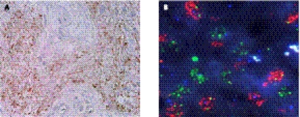
Figure 2.
Molecular heterogeneity in glioblastoma. (A) Immunohistochemistry for the mutant EGFR receptor EGFRvIII demonstrates a heterogeneous staining pattern within the tumor. Images from Nishikawa et al. (2004) used with permission. (B) Multicolor FISH reveals distinct subpopulations of either EGFR (red) or PDGFRA (green) amplification within a glioblastoma specimen. Images obtained from Cameron Brennan.

