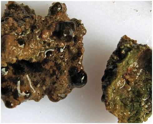
Figure 1.
Photograph of the Ward Hunt Lake microbial mats. The mats were photographed 30 min after sampling, on July 7, 2010. The mat on the left shows the view from the top, with black colonies distributed over the pink surface. The mat on the right has been turned upside down to show the green layer, which extended down into interstices of the underlying rocky substrate. The black colonies were up to 5 mm in diameter.

