
Figure 4.
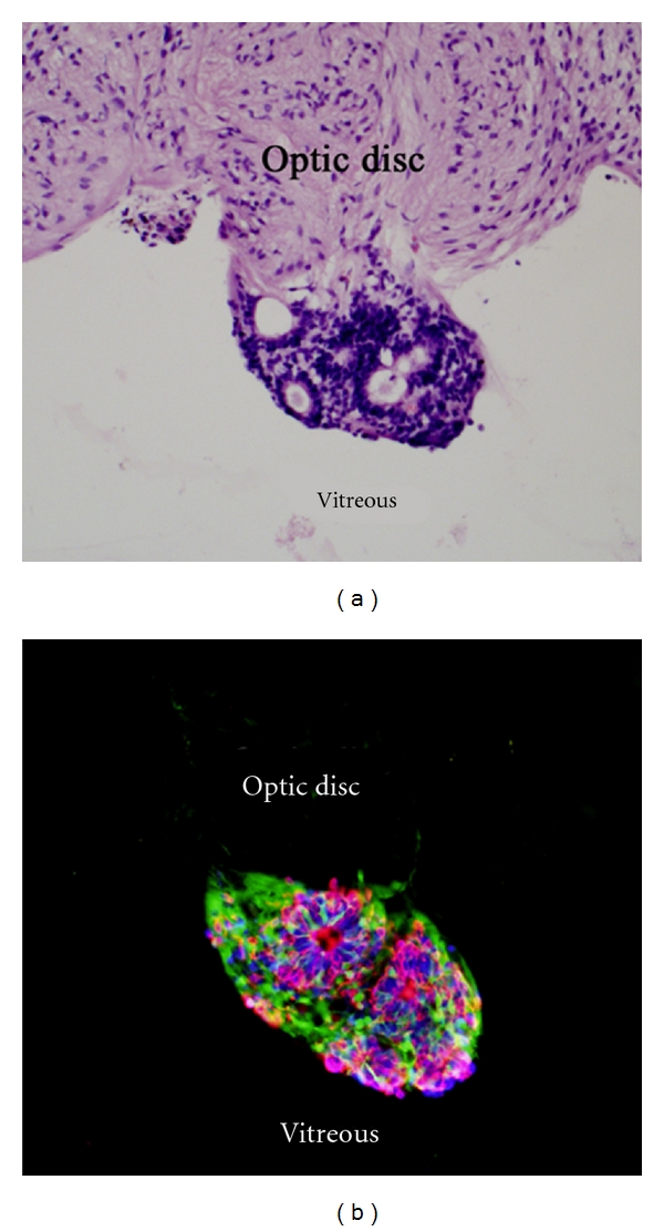
Grafted cells form rosettes and express photoreceptor markers. Recipient was 6 weeks of age at time of transplantation and experiment terminated 5 weeks later. The H&E section (a) shows a cluster of grafted cells adhering to vitreal surface of the host optic nerve head, likely a result of reflux around the time of surgery. This cluster contains multiple rosettes of the type formed by photoreceptor cells, with darkly staining nuclei densely packed in a peripheral rim and lighter staining material in the rosette's core. Fluorescence imaging (b) confirms the cluster to be GFP+ and therefore of donor origin. Immunolabeling for rhodopsin (red) and recoverin (blue) demonstrates photoreceptor marker expression by a subset of grafted cells. Cells expressing photoreceptor markers are predominantly found within rosettes.