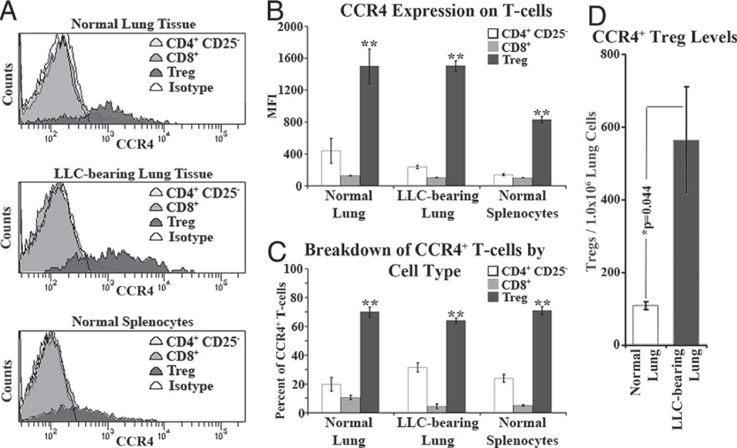
FIGURE 2.
CCR4 expression on Tregs is unique among T cells. Homogenates from both normal and LLC-bearing lung tissue along with normal splenocytes were immunofluorescently stained for CD8, CD4, CD25, FoxP3, and CCR4. A, Representative histograms of CCR4 expression among T cell types. B, Across all types of homogenate, Tregs had significantly higher MFI of CCR4 staining compared with both CD8+ and CD4+CD25− cells (**, p < 0.05 vs CD8+ and CD4+CD25−). Similarly, of all CCR4+ T cells in the homogenates, Tregs comprised a greater percentage as compared with the CD8+ and CD4+CD25− populations (C). In addition, LLC-bearing tissue contains a significantly increased number of CCR4+ Tregs (D).

