
Fig. 4.
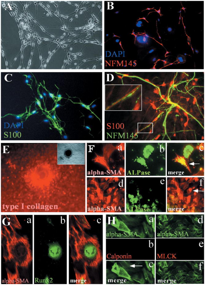
Differentiation potentials of CNC cells in culture. Sorted CNC cells are maintained in MCDM for 5 days and are switched to DMCDM medium for additional 4 days. The differentiation status of cultured CNC cells is evaluated by using different markers. A: Some of the neural crest–derived cells display typical neural cell morphology following the switch of cell culture medium from MCDM to DMCDM. B: Double staining with antibodies against NF145 (red) and DAPI (blue). C: Double staining with antibodies against S100 (green) and DAPI (blue). D: Double staining with antibodies against NF145 (green) and S100 (red). Part of the axon is enlarged to show the parallel alignment of neuron and glial cell (inset), faithfully mimicking the relationship in vivo. E: Calcified nodule is stained with antibody against type I collagen (red). Inset at top left corner is the same calcified nodule under light microscope and is positive for osteopontin expression, therefore, validating the differentiation of CNC cells into osteoblasts. F: Double staining with ALPase (green) and α-SMA (red) antibodies indicates that some CNC cells express both alpha-SMA and ALPase (F, c: arrow). While some CNC cells only express alpha-SMA (F, f: arrow), most of ALPase positive cells are also alpha-SMA positive (F, f: green and red staining merged as yellow). G: Double staining with α-SMA (G, a, red) and RUNX2 (G, b, green) antibodies indicates that some CNC cells express both alpha-SMA and RUNX2 (G, c, merged a and b). H: Double staining with α-SMA + calponin antibodies (a–c) or †-SMA + MLCK (d–f) show that these α-SMA positive cells do not express calponin or MLCK.