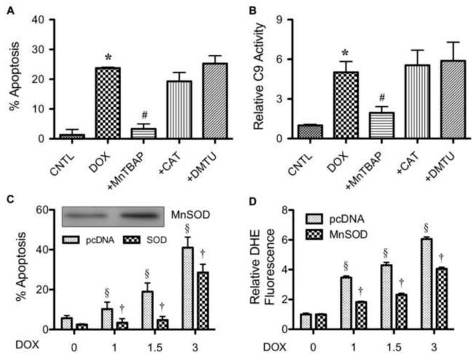
FIGURE 4.
Superoxide mediates doxorubicin-induced apoptosis. A, HaCaT cells were pretreated for 30 min with MnTBAP (50 μM), catalase (CAT, 7,500 units/ml), or DMTU (5 mM) followed by DOX treatment (1.5 μM) for 24 h and analyzed for apoptosis by Hoechst 33342 assay. B, cells were similarly treated with the ROS scavengers and DOX as described above and analyzed for caspase-9 activity using the fluorometric substrate FAM-LEHD-fmk. C, cells were transiently transfected with mitochondrial superoxide scavenging enzyme MnSOD or control pcDNA3 plasmid as described under Material and Methods. Transfected cells were treated with DOX (0-3 μM) for 24 h and analyzed for apoptosis by Hoechst 33342 assay. MnSOD expression of the transfected cells were analyzed by Western blotting. D, transfected cells were similarly treated with DOX and analyzed for superoxide generation by flow cytometry at 2 h after the treatment. Plots are mean ± S.D. (n = 3). *, p < 0.05 versus non-treated control. #, p < 0.05 versus DOX-treated control. §, p < 0.05 versus vector-transfected cells. †, p < 0.05 versus DOX-treated vector-transfected cells.

