
Figure 6. Cell migration from the dorsal sclerotome is unaffected in Myf5-Cre;Tgfbr2flox/flox mice.
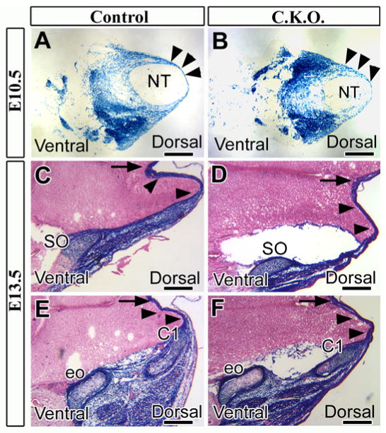
Cross sections stained for lacZ at E10.5 (A, B) and E13.5 (C-F). (A, B) Control (A: Myf5-Cre;Tgfbr2flox/+;R26Rflox/+) and conditional knockout (B: Myf5-Cre;Tgfbr2flox/flox;R26Rflox/+) mice contained mesoderm-derived cells (dark blue) migrating into the mid-dorsal area (black arrowheads). (C-F) Mesoderm-derived cells (black arrowheads) migrate to the mid-dorsal region in both control and conditional knockout mice, arriving at the supraoccipital bone (SO) and the neural arch of C1 vertebra (C1). Black arrows indicate the dorsal midline. Abbreviations: NT, neural tube; eo, exoccipital bone. Scale bars: 500 μm in A-F.