
Figure 9. The pattern of cell proliferation is altered in the supraoccipital bone primordium of Myf5-Cre;Tgfbr2flox/flox mice.
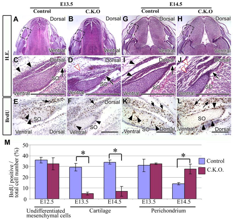
Cross sections of E13.5 (A-F) or E14.5 (G-L) supraoccipital bone primordium stained with hematoxylin and eosin (A-D, G-J) or BrdU (E, F, K, L). (C-F, I-L) Enlarged areas from the dashed black boxes of A, B, G, and H are shown in C/E, D/F, I/K, and J/L, respectively.
(A, C) Control mice have well-organized perichondrium (C, black arrow) and muscle tissue (C, black arrowheads). (B, D) The perichondrium of conditional knockout mice is organized (D, black arrow), but muscle tissue is disorganized (D, white arrowhead). (E, F) Control and conditional knockout mice contain BrdU positive cells in the cartilage and the perichondrium of the supraoccipital bone (black arrowheads) and in the muscle cells (black arrow). (G, I) The perichondrium (I, black arrow) and muscle tissue (I, black arrowheads) are well-organized in control mice. (H, J) Conditional knockout mice have a well-organized perichondrium (black arrow), but disorganized muscle tissue (J, white arrowheads). (K, L) In control mice, BrdU positive cells are present in both the cartilage and perichondrium of the supraoccipital bone (black arrowheads) and also in the muscle (black arrows). BrdU positive cells are visible in the muscle (black arrows) and perichondrium (black arrowheads) of the supraoccipital bone in conditional knockout mice, but not in the cartilage of supraoccipital bone. (M) Statistical analysis of cell proliferation activity in control and conditional knockout mice. Five randomly selected, non-overlapping samples were used to obtain the BrdU labeling index from each experimental group. Student t-tests were used for statistical analysis. *:P<0.05. Abbreviations: SO, the primordium supraoccipital bone. Scale bars: 300 μm in A-L.