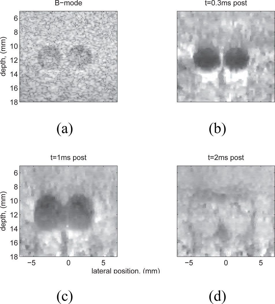
Fig. 2.
Experimental data: matched B-mode (a) and normalized ARFI displacement images, (b)–(d), of a Computerized Imaging Reference Systems, Inc. (CIRS, Norfolk, VA) custom tissue mimicking phantom (E=4 kPa) with two 3 mm spherical lesions (E=58kPa). The lesion contrast in the ARFI images is largest at t=0.3 ms (b), decreases with time after excitation (c), and reverses later in time (d). In addition, the lesion size appears to grow with time post-force, which is caused by shear wave propagation and reflection at lesion boundaries (35). Note also the ’posterior enhancement’, or increase of displacement beneath the lesions (b), (c). This is because the lesions were slightly less attenuating than the surrounding tissue, thus the tissue beneath the lesions experienced larger radiation force than that adjacent to it. Figure reproduced with permission from:(36)

