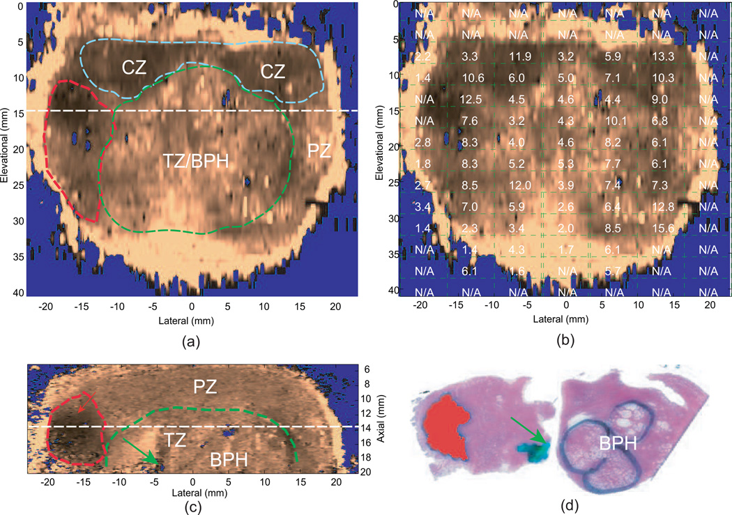
Fig. 5.
Qualitative ARFI images with the co-registered reconstructed shear moduli and matched histological slides from an ex vivo prostate specimen. Images were obtained with a linear array mounted on a translation stage used to interrogate the entire 3D prostate. The central zone (CZ), transition zone with benign prostatic hyperplasia (TZ/BPH) and prostate cancer (PCa) are circled in blue, green and red dashed lines in the ARFI images, respectively. The urethra is indicated by the green arrow. (a) coronal ARFI image with zonal anatomy, PCa and BPH indicated; (b) coronal ARFI image with co-registered reconstructed shear moduli overlaid; (c) axial ARFI image with zonal anatomy, PCa and BPH indicated; (d) co-registered histological slide of the axial ARFI image, in which PCa is masked in red and BPH is circled in black. (a) and (c) are perpendicular to each other and intersect at the dashed lines. These data were obtained with a VF105 linear array mounted to a mechanical translation stage and a Siemens Antares scanner. Figure reproduced with permission from:(46)

