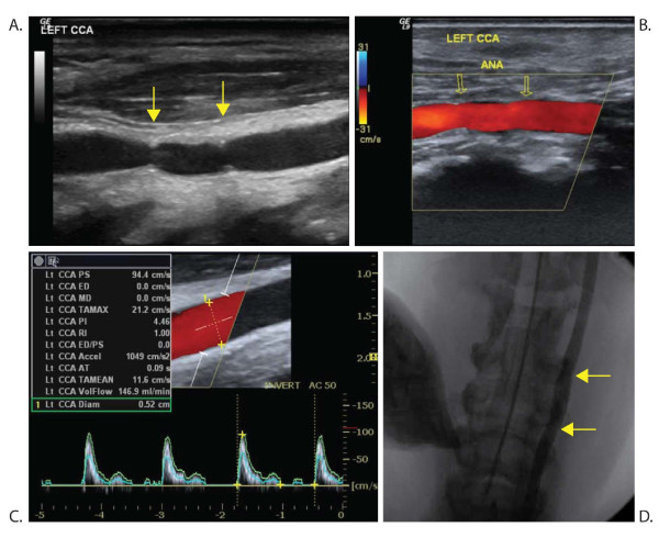
Figure 2.
Ultrasound and angiography of a common carotid artery with vein graft. Gray-scale ultrasound (A) and angiography (D) showing the anatomy. Color Doppler (B) and spectral Doppler (C) investigation showing normal flow. Yellow arrows point at the anastomoses (ANA). CCA = common carotid artery. PI = pulsatility index. RI = resistive index.

