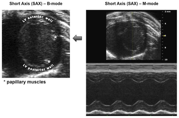
Figure 1. Echocardiographic landmarks.
B-mode image of the left ventricle (LV) short axis (SAX) and resulting M-mode image showing the projection obtained for measurement of stroke volume and other parameters. In particular, note the position of the mitral valve papillary muscles at 2 and 4 o’clock used to establish correct and consistent transducer positioning.

