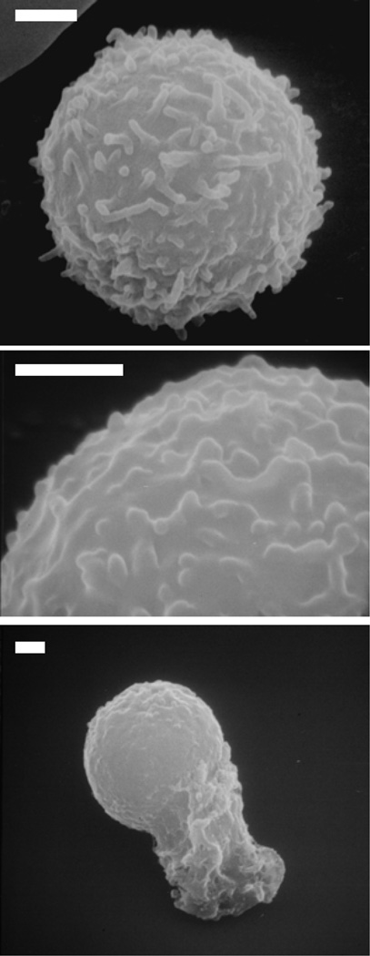
Figure 3. Scanning electron micrographs of human leukocytes.
The surfaces of human leukocytes are completely covered by sub-micron membrane folds (microvilli) that are on the order of 0.2µm in height. The length and shape of the membrane folds are non-uniform. The top micrograph shows a quiescent leukocyte whereas the bottom image shows an activated cell. The middle image shows the microvilli in more details. Bar = 1µm.

