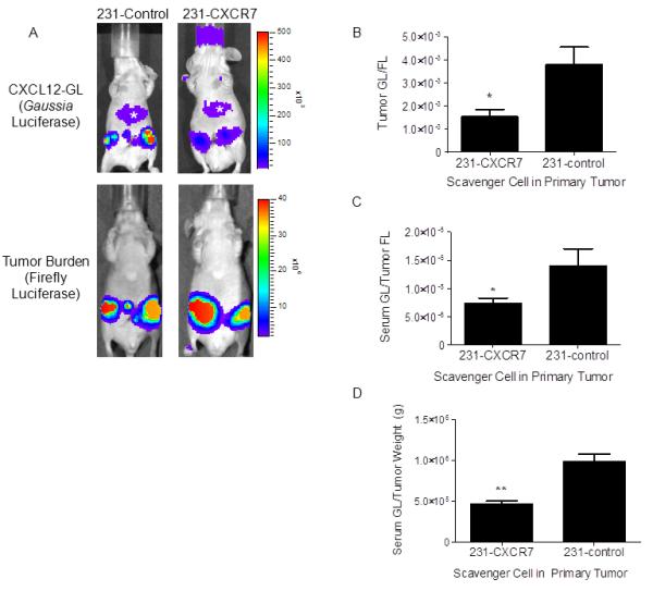
Figure 2. CXCR7 reduces CXCL12 in primary breast tumors.

A) Mice were implanted with orthotopic breast tumor xenografts of 231-CXCR7 or 231-control cells with HT1080-CXCL12-GL/FL cells. Representative images are presented from Gaussia and firefly luciferase bioluminescence imaging for tumors with 231-CXCR7 or 231-control cells. Asterisk denotes bioluminescence from oxidation of coelenterazine in liver. Scale bar depicts range of photon flux values as pseudocolor display with red and blue representing high and low values, respectively. B) Photon flux from CXCL12-GL was normalized to firefly luciferase in each tumor and presented as mean values + SEM (n = 5 per group). C) CXCL12-GL in serum was quantified by bioluminescence. Data were normalized to photon flux from firefly luciferase in each mouse. D) Bioluminescence from CXCL12-GL in serum was normalized to weight of excised tumors. Data were graphed as mean values + SEM. *, p < 0.05; **, p < 0.01.
