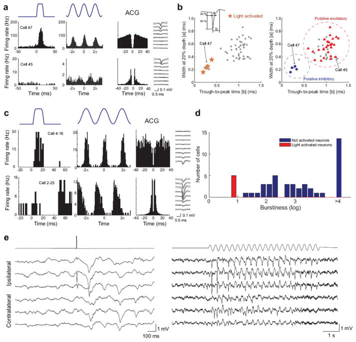
Figure 7.

In vivo identification of light-activated neurons in the hippocampus and thalamus of Pvalb-IRES-Cre;Ai32 mice. (a) Excitation of ChR2-expressing neurons in the hippocampal CA1 region during waking state. Top row shows peristimulus histogram of a Pvalb+ neuron (Cell 47) transiently activated by single pulses or 8-Hz sinus light stimulation (1 mW). Also shown are the autocorrelogram (ACG) and waveform (± s.e.) of the neuron. Note typical ACG for a fast firing putative basket cell 35. Bottom row shows the same arrangement for a nearby pyramidal cell (Cell 45). Note ACG typical of bursting neurons. (b) Optogenetic (left) and physiological (right) classifications of neuron types in the hippocampus strongly overlap. Physiological segregation of simultaneously recorded neurons is based on two parameters – spike width and trough-to-peak time (inset). (c) Activation of ChR2-expressing neurons in the thalamus during anesthesia. Top row shows a reticular nucleus Pvalb+ neuron (Cell 4-16) in response to single pulses or 10-Hz sinus light stimulation. Bottom row shows a simultaneously recorded thalamocortical neuron (Cell 2-23) with typical bursting pattern in ACG 36, 37. (d) Distribution of burst index in the activated (putative reticular, red) and suppressed (putative thalamocortical, blue) neurons. Burst index is the ratio of spikes with short (<6 ms) inter-spike intervals relative to other spikes in the same session. (e) Light-evoked cortical patterns in response to reticular nucleus stimulation in the waking Pvalb-IRES-Cre;Ai32 mice. Single pulse (left) and sinus pattern (10 Hz, right) evoked activities are epidural recordings from the ipsilateral and contralateral parietal areas.
