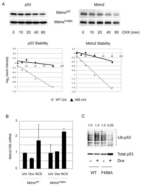
Figure 2. Stabilization of p53 upon MdmxF488A expression is post-translational.
(A) Following induction of MdmxWT or MdmxF488A, cycloheximide was added for the indicated times prior to blotting for p53 and Mdm2. Band intensities were calculated using the LiCor/Odyssey image analysis system, and plotted as log2 values. (B) Mdm2 mRNA was analyzed by qPCR following addition of Dox, or the damaging agent NCS (300 ng/ml) as a positive control. (C) Cells were transfected with Histidine-tagged ubiquitin and doxycycline was added to induce MdmxWT or MdmxF488A. After 24h, cells were lyzed and ubiquitylated proteins were pulled down using Ni2+-agarose beads. Following SDS-PAGE, p53 was detected using DO-1. Numbers above each lane represent the ratio of ubiquitylated p53 species to the total amount of p53 in the lysate.

