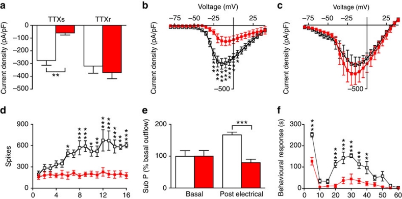
Figure 5. Reduced electrically evoked wind-up and substance P release in the spinal cord of Nav1.7Advill mice.
Littermate (white columns/boxes), Nav1.7Advill (red columns/boxes) N shown as littermate/Nav1.7Advill. (a) TTX-sensitive and TTX-resistant inward sodium currents recorded from Nav1.7Advill DRG neurons (N=11/6) Current–voltage relationships for sodium currents in littermate control (N=11) and Nav1.7Advill DRG neurons (N=6) in the presence (b) or absence (c) of 500 nM TTX. (d) WDR spinal recording from Nav1.7Advill and littermate mice in response to electrical stimulation of the sciatic nerve (N=6 per group). (e) Electrically evoked substance P release into the dorsal horn measured by radioimmunoassay in Nav1.7Advill and littermate mice (N=7/8). (f) First and second phase responses observed in Nav1.7Advill mice after intraplantar injection of 20 μl of 5% formalin (N=6/5). All data analysed by two-way analysis of variance followed by the Bonferroni post hoc test. Results are presented as mean±s.e.m. *P<0.05, **P<0.01 and ***P<0.001 (individual points).

