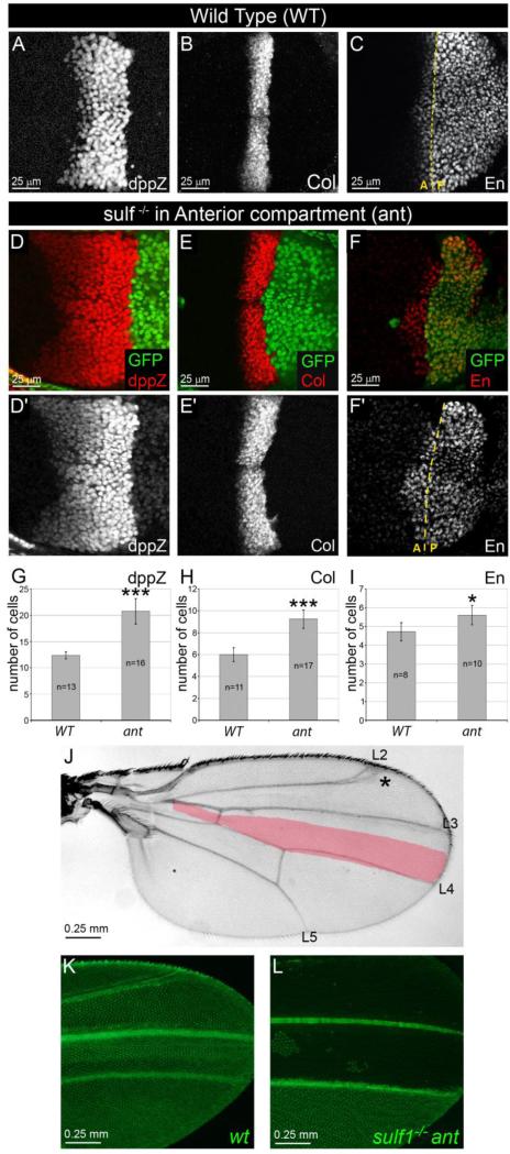
Figure 2. Removal of DSulf1 activity in Hh receiving compartment results in Hh gain-of-function phenotype.
(A-F’) Expression patterns of dppZ (A, D and D’), Col (B, E and E’) and En (C, F and F’) in wt (A-C) and in Dsulf1ΔP1 mitotic clones restricted to the A compartment (D-F’) as visualised by the absence of GFP staining (green). Note the anterior extension of dpp (D, D’), Col (E, E’) and En (F, F’) expression domains compared to wt (A, B and C, respectively). (G-I) Number of cell rows expressing dppZ (G), Col (H) and En (I) in wt and in wing discs containing anterior (ant) Dsulf1ΔP1 clones. Error bars represent the standard deviation (*=P<0.05; ***=P<0.0005 using a t-test). (J) Adult wing carrying anterior mutant clones showing a broadening of the L3-L4 spacing (40±3 trichomes) compared to wt (30±1 trichomes, red in J). Note additional defects affecting the positioning of the L2 vein (asterisk). (K-L) Visualization of GFP in wings obtained from young emerging adults. Note the broadening of the L3-L4 spacing in wing containing Dsulf1ΔP1 mutant cells in the A compartment (L) compared to wt (K).

